


(281) 713-2962
800 Rockmead Drive, Suite 155
Kingwood, TX 77339
[email protected]
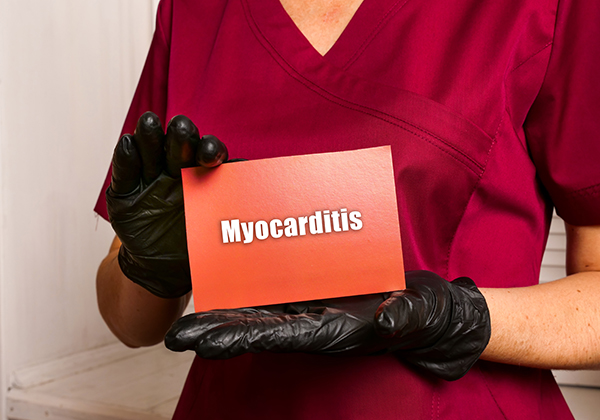
Foundation for Sarcoidosis Research Advanced Cures Registry (FSR-SARC Registry)
Status: Recruiting
Location: Foundation for Sarcoidosis Research
Conditions: Foundation for Sarcoidosis Research
City/State:
Chicago, Illinois
Contact Information:
Leslie Serhuck, MD MA Mbioethics
312-341-0500
[email protected]
Tricha Shivas, MBe
312-341-0500
[email protected]
Brief Summary:
The goal of the study is to create a longitudinal study of patient reported outcomes for people living with sarcoidosis that maintains privacy. Patients report on the following: demographics, disease symptoms, diagnostic journey, provider experience, disease treatment, and burden of disease. The goal is to create a natural history of sarcoidosis, support research, and better understand the needs of the sarcoidosis community.
Detailed Description:
Participants review a document, Understanding Your Participation, and check boxes on the Participant Informed Consent document that confirms they understand the risks/benefits of participation (or Assent if the patient is a minor age 7-18), they create an online account, and then are asked to complete the baseline survey questionnaire. Participants confirm they understand that their participation is completely voluntary, that their identifying information will be secured and encrypted, their private health information will be stored separately in a secure database. Their private information will never be shared with other people, unless its required by law. The registry may share de-identified information with researchers and other databases. Their personal information will be protected and not shared. They may choose to stop their participation at any time by contacting FSR. They are not required to fill out all the questions and can leave any unanswered. They will be contacted by the registry once a year to update or correct their health information. They can choose to be contacted by FSR if a study becomes available that they may wish to know more about.
Rheumatology Patient Registry and Biorepository
Status: Recruiting
Location: Yale-New Haven Hospital
Conditions: Yale-New Haven Hospital
City/State:
New Haven, Connecticut
Contact Information:
Nicolas Page
203-737-5571
[email protected]
Brief Summary:
To facilitate clinical, basic science, and translational research projects involving the study of rheumatic diseases.
Detailed Description:
A rheumatology biorepository will be created to permit comparative analyses between the rheumatic diseases in order to increase the understanding of disease pathogenesis. Patients seen at Yale clinics diagnosed with rheumatic diseases are invited to participate in this study. These rheumatic diseases include, but are not limited to: adult onset Still’s disease, ankylosing spondylitis, psoriatic arthritis, reactive arthritis, antiphospholipid syndrome, systemic lupus erythematosus, Behcet’s disease, dermatomyositis, polymyositis, giant cell arteritis and other vasculitides, Lyme’s disease, mixed connective tissue disease, polymyalgia rheumatica, rheumatoid arthritis, sarcoidosis, systemic sclerosis (scleroderma), Sjogren’s syndrome, and undifferentiated connective tissue disease.
A Study to Evaluate the Efficacy, Safety, and Tolerability of BMS-986278 in Participants With Progressive Pulmonary Fibrosis
Status: Recruiting
Location: Stanford Hospital and Clinics, University Hospitals Cleveland Medical Center
Conditions: Stanford Hospital and Clinics, University Hospitals Cleveland Medical Center
City/State:
Stanford, California
Cleveland, Ohio
Contact Information:
BMS Study Connect Contact Center www.BMSStudyConnect.com
855-907-3286
[email protected]
Brief Summary:
The purpose of this study is to evaluate the efficacy, safety, and tolerability of BMS-986278 in Participants with Progressive Pulmonary Fibrosis.
Interstitial Lung Disease Research Unit Biobank
Status: Recruiting
Location: University of Kansas Medical Center
Conditions: University of Kansas Medical Center
City/State:
Kansas City, Kansas
Contact Information:
Kimberly Lovell, Ph.D.
913-588-6067
[email protected]
Brief Summary:
Establish a interstitial lung disease (ILD) registry and biorepository to lead towards a further understanding of the disease.
Detailed Description:
The University of Kansas ILD and Rare Lung Disease clinic sees hundreds of new patients per year. The investigators would like to leverage this resource to develop an Interstitial Lung Disease Research Unit (ILDRU) repository and database to help develop new methods for early diagnosis, uncover underlying genetic and environmental risk factors, as well as potential treatment targets in the broad range of interstitial lung diseases and rare lung diseases (RLD).
A Study of the Natural Progression of Interstitial Lung Disease
Status: Recruiting
Location: University of Chicago
Conditions: University of Chicago
City/State:
Chicago, Illinois
Contact Information:
Spring Maleckar
773-834-4053
Brief Summary:
We propose to acquire data and blood samples on all patients being cared for by the Interstitial Lung Disease (ILD) program. Additionally, we will collect data and blood samples from a control group for comparator purposes. In doing so, we will be able to describe the “phenotypic” expression of these diseases.
Inhaled Treprostinil in Sarcoidosis Patients With Pulmonary Hypertension
Status: Recruiting
Location: University of Florida
Conditions: University of Florida
City/State:
Gainesville, Florida
Contact Information:
Christina M Eagan, DNP
352-273-8990
[email protected]
Name: Ali Ataya, RN
Phone Number: 352-273-8740
Email: [email protected]
Brief Summary:
This study aims to evaluate the efficacy and safety of inhaled treprostinil in subjects with sarcoidosis-associated interstitial lung disease and pulmonary hypertension.
Detailed Description:
Pulmonary sarcoidosis-associated pulmonary hypertension is classified as WHO Group 5 pulmonary hypertension and may occur in anywhere from 5-20% of sarcoidosis patients. Inhaled treprostinil has shown clinical improvements in exercise capacity after 12 weeks of therapy in patients with WHO Group 1 pulmonary hypertension. More recently, there has been interest in using inhaled PAH-specific therapies for the treatment of pulmonary hypertension associated with interstitial lung disease.
The investigators believe that those patients with pulmonary hypertension in the setting of sarcoidosis-associated interstitial lung disease are a unique population which may potentially benefit from inhaled, targeted pulmonary arterial hypertension therapy (inhaled treprostinil) while minimizing the adverse effects associated with systemic pulmonary vasodilators. This study aims to evaluate the efficacy and safety of inhaled treprostinil in subjects with sarcoidosis-associated interstitial lung disease and pulmonary hypertension.
Diagnostic Yield of Intranodal Forceps Biopsies in Mediastinal Adenopathy
Status: Recruiting
Location: The George Washington University Hospital
Conditions: The George Washington University Hospital
City/State:
Washington D.C.
Contact Information:
Khalil Diab, MD
2027412180
[email protected]
Benjamin Delprete, DO
2027412180
[email protected]
Brief Summary:
The investigators will compare endobronchial ultrasound transbronchial needle aspiration (EBUS-TBNA) with intranodal forceps biopsy (EBUS-IFB) as it relates to the rate of diagnosis of suspected sarcoidosis.
Detailed Description:
This is prospective, single center randomized comparative study to determine the diagnostic yield and specimen quality of endobronchial ultrasound guided intranodal forceps biopsy of patients with suspected sarcoidosis based solely on imaging. This will be a single group study and will compare transbronchial needle aspiration via 19 or 21-gauge needle with intranodal forceps biopsy.
The study aims to answer a knowledge gap a as to whether the diagnostic yield and specimen quality of EBUS-TBNA with a 19G needle is less than those obtained by 1.9mm or greater intranodal forceps biopsy. The study proposed here will add to the field by further elucidating whether this procedure is beneficial for the diagnosis as it pertains to suspected sarcoidosis.
The anticipated required enrollment is 55 patients to achieve an α of 0.05 and β of 0.2. This assumes an unassisted diagnostic yield of 62.5% with standard of care EBUS-TBNA as reported in Ray et al, and a diagnostic supplementation to 80% yield with intranodal forceps biopsies.
Full-Field Optical Coherence Tomography (FFOCT) for Evaluation of Bronchoscopic Small Biopsy Specimens
Status: Recruiting
Location: Johns Hopkins University
Conditions: Johns Hopkins University
City/State:
Baltimore, Maryland
Contact Information:
Jeffrey Thiboutot, MD
410-502-2533
[email protected]
Brief Summary:
This study sets out to register imaging of small biopsy specimens obtained during bronchoscopy using full-field optical coherence tomography against standard histologic evaluation.
Detailed Description:
Small biopsy specimens obtained through bronchoscopy are commonly employed for the diagnosis and staging of thoracic malignancies. Diagnostic yield is dependent on tissue quality and quantity in specimens obtained through bronchoscopy, and it is thus important to ensure adequate sampling. Rapid on-site cytology (ROSE) is a method used by having a cytotechnologist at the bedside to prepare and analyze specimens to improve the quality of tissue acquisition during bronchoscopy. Although effective, ROSE expertise is not always available to proceduralists, is costly, and reproducible techniques that can be deployed across multiple tiers of institutions are needed across the globe.
Optical coherence tomography (OCT) is an emerging technique which may provide real-time imaging with resolution approaching that of typical histopathology. This has several benefits over ROSE using histopathologic evaluation including rapid imaging with minimal tissue processing, preservation of tissue specimens for molecular testing, enhanced intracellular contrast, and adaptation to machine learning approaches to allow for a reproducible and consistent result. In fact, full-field OCT has recently been applied in several tissue types for evaluation of adequacy of pathologic specimens and evaluation of malignancy, among others. To the best of the investigators’ knowledge, this technology has not yet been evaluated for assessment of specimen quality in bronchoscopic procedures.
Thus, the investigators propose a study of full-field OCT for evaluation of small biopsy specimens obtained through bronchoscopy. The investigators aim to demonstrate the feasibility of this technology in the workflow of bronchoscopy and compare to current evaluation methods including rapid on-site evaluation (ROSE). ROSE is commonly used to evaluate adequacy of tissue diagnosis during bronchoscopic procedures including at this institution. However, studies have not shown definitive benefits over bronchoscopy without ROSE, and current expert guidelines suggest bronchoscopy with Endobronchial Ultrasound (EBUS) transbronchial needle aspiration (TBNA) may be performed with or without ROSE.
Full-field OCT has several potential benefits compared to ROSE, including rapid analysis with minimal tissue processing and preservation of tissue for further molecular testing. In addition, OCT has been used to assess surgical biopsy specimens in a non-destructive manner, so the tissues can be analyzed after imaging using standard cytological and pathological methods. Full-field OCT evaluation may be applied to other diseases in addition to further augmenting the diagnostic ability through the use of machine learning approaches.
Read more
Medication Adherence in Patients With Sarcoidosis
Status: Recruiting
Location: Johns Hopkins Bayview Asthma and Allergy Center, Johns Hopkins Greenspring Station
Conditions: Johns Hopkins Bayview Asthma and Allergy Center, Johns Hopkins Greenspring Station
City/State:
Baltimore, Maryland
Timonium, Maryland
Contact Information:
Michelle Sharp, MD, MHS
410-550-7753
[email protected]
Amanda Sevilla, BA
410-550-1859
[email protected]
Brief Summary:
The goal of the study is to look at the relationship between how individuals with Sarcoidosis take the sarcoidosis medicines and how it affects the disease, to evaluate any factors that may make individuals not want to take the medicines, and to develop and refine ways to help support individuals with Sarcoidosis especially when it comes to the medicines. The overall hypothesis is higher medication adherence will be associated with better clinical outcomes in sarcoidosis. The investigators will enroll 150 patients with biopsy proven pulmonary sarcoidosis for at least one year who are on any oral treatment regimen for at least six months into a 12-month longitudinal study.
A Study of XTMAB-16 in Patients With Pulmonary Sarcoidosis
Status: Recruiting
Location: Xentria Investigative Site
Conditions: Xentria Investigative Site
City/State:
Greenville, North Carolina
Birmingham, Alabama
Denver, Colorado
Jacksonville, Florida
Chicago, Illinois
Iowa City, Iowa
Albany, New York
New York, New York
Philadelphia, Pennsylvania
Baltimore, Maryland
Minneapolis, Minnesota
Detroit, Michigan
Cincinatti, Ohio
Charleston, South Carolina
Houston, Texas
Charlottesville, Virginia
Contact Information:
Xentria, Inc.
224-443-4615
[email protected]